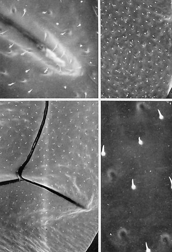
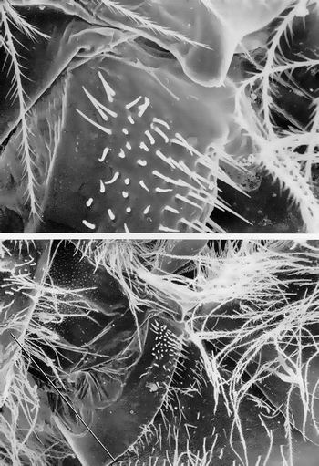
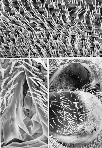
DRONEWING,VENTRALSURFACE
BOTTOMLEFT.Hindwing,ventralview,atlowmagnification.Severalveinsintersectinthisfield.(x90)
TOPLEFTVentralview,nearthetipofhindwing.Exceedinglysmall,cuticularspinesrandomlyariseinthisarea.Somescatteredpitsarealsovisible.(x420)
TOPRIGHT.Forewingtip,ventralview.Numerousscatteredsmall,bluntcuticularspinesarefoundonthissurface.Thepitsrepresentthebasesoftheshortcounterpartspinesarisingonthereversesurfaceofthiswing.(x130)
BOTTOMRIGHT.Highermagnificationoftheventralforewinginthetoprightmicrograph.Thespinedensityis1/900microm^2.(x750)
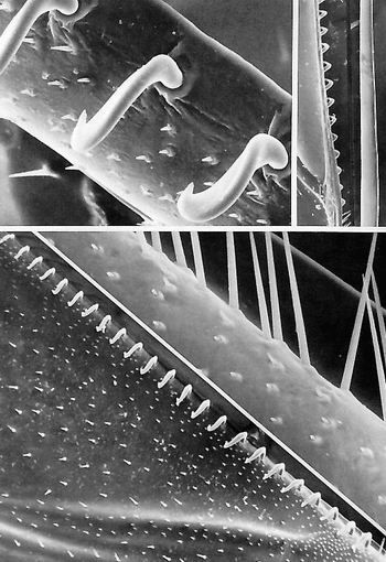
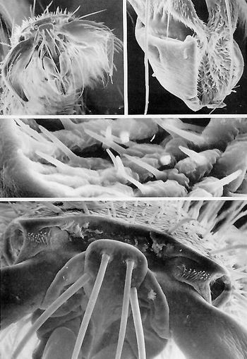
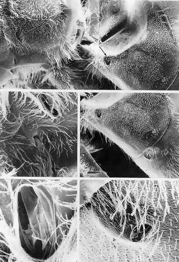
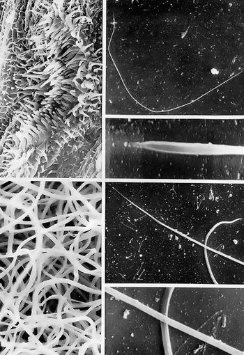
DRONEWINGHOOKS
BOTTOMLEFT.LeadingedgeofthehindwingThehooksengagetheposteriormarginoftheforewingonlyduringflight,whentheforewingandhindwingbeatasone.Microtrichiaareabundantoverthewingsurface.(x136)
BOTTOMRIGHT.Non-hook-bearingportionofthehindwinginthebottomleftmicrograph.Socketedhairs,bothlongandveryshort,projectfromthisvein.(x800)
TOPRIGHT.Photorriontageoftheventralforewinghindmargin,aroundwhichthehooksengage,andthehookedleadingedgeofthehindwing.(x110)
TOPLEFT.Close-upofthewinghooks.Thesearesomewhatlargerthanthoseoftheworker.Socketedsmallpegorgansareabundantonthesurfaceengagedbythetrailingedgeoftheforewing.(x720)
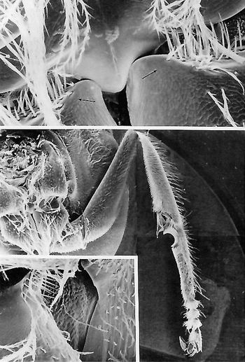
DRONEFORELEG
MIDDLE.Photomontageoftheentireleftforeleg.Proximallytodistally,afterthecoxa(firstlegsegmentfromthebody)arethetrochanter,femur,tibia,basitarsus(withantennacleanernotch),threeshorttarsalsegments,pretarsus,andclaws.(x31)
TOP.Prothorax,ventralview.Twoscaledcoxae(medioanteriorsurfaces)abutoneithersidetoasmooth-surfacedbasisternum.Mechanoreceptorhairplatesofthecoxaeengagethelateralbasisternum(arrows).(x168)
BOTTOMLEFTLateraledgeofthecoxaextendingdiagonallythroughthecenterofthefield.Amechanoreceptorhairplateonthelateroanteriorsurfaceengagestheadjacentsternum.(x93)
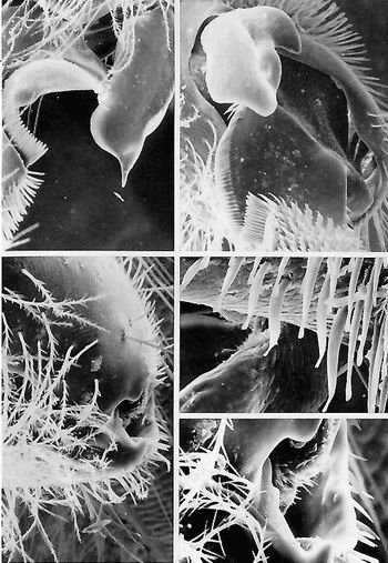
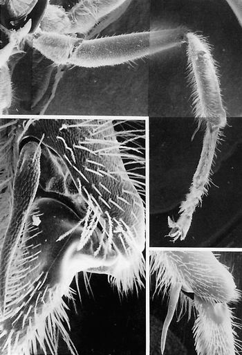
DRONEFORELEG,CLOSE-UP
TOPLEFT.Tibia-basitarsusjoint,theregionoftheantennacleaningapparatus(comb).Thefibula(upperright)isaclasplikespurthatclosesoverthesemicirculartoothednotch(rimmedwithcomblikehairs).Theantennaisinsertedinthisenclosureanddrawnupwardtocleandebrisfromitscuticularreceptors.Thefibulafitsintotheproximalportionofthebasitarsustoformanembracearoundallantennasurfaces.(x161)
TOPRIGHT.Dorsalviewofthefibulashowingthenotchoftheantennacleaner.Fromthisangleitisapparentthatthefibulaiscontouredintoseverallobestomakebettercontactwiththecircular(incrosssection)antenna.(x195)
MIDDLERIGHTHighermagnificationofthetibia-basitarsusjoint.Apatchofunusualcuticularsetaeisonthetibia(seethetoprightmicrograph).Thesesetaehavetwistedblunttipswithseveralparallelridgesalongthelongaxisofthehair.(x496)
BOTTOMRIGHT.Femur-tibiajoint.Theintersegmentalmembraneisstuddedwithshort,acutecuticularspines.(x770)
BOTTOMLEFTFemur-tibiajoint.Thetibiaisveryflexedrelativetothefemur.Thelong,straight,unbranchedhairsofthecompoundeyeareinthefarbackground.(x280)
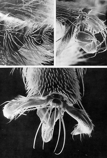
DRONEFOREFOOTANDHINDFOOT
TOPLEFT.Surveyofthehindfoot.Theclawsaresimple,unlobed,hollowhooksfringedwith(probablysensory)hairs.(x100)
TOPRIGHT.Arolium(fromthehindfoot)showingitshirsutemedialsurfaceandglabrous,groovedexterior(comparewithPlate1.34,thequeenforefootandhindfoot).Threelongspineshangdownfromthemedianscleriteabove.(x400)
MIDDLE.Highermagnificationofthehairsliningthemedialsurfaceofthearolium.(x4,250)
BOTTOM.Forefoot(prothoracicfoot).Intheforegroundisthemediansclerite,fromwhichextendfourstout,slightlycurvedspines.Thebasalportionsoftherightandleftclawsareoneithersideofthemediansclerite.Theventrodistalmarginofthefifthtarsalsegmentarisesovertheclawbasesandmediansclerite.Leftandrightsensoryhairplatesareevidentabovetheclaws;whentheclawsareraisedtheycontacttheseputativemechanoreceptorstoinformthedronewhetheritsclawsareextendedorflexed.(x510)
DRONEMIDDLELEG
TOP.Photomontageoftheleftmiddle(mesothoracic)leg.Thecoxaisobscuredbut,proximallytodistally,thetrochanter,femurtibia(withspineorwaxspurpointeddownward),basitarsusandthreeadditionaltarsomeres,andthepretarsuswithclawsarevisible.(x31)
BOTTOMRIGHT.tibia-basitarsusJoint.Thisisamonocondylic(singlepoint)articulation.Therelativelylargesizeandstoutnessofthetibialspineareevident.(x93)
BOTTOMLEFT.Tibia-basitarsusJoint.Thisviewandmagnificationmakeapparentthedifferencesinthecuticularsurfacesbetweenthescaleliketibialbase,thepebble-grainedtibialspine,andthesmoothheadofthebasitarsus.Thetwoformsofsetae(branchedandsimpletrichoid)onthetibiaarevisibleatthisarticulation,aswellasthescalesonthemtersegmentalmembraneatthisJoint.(x355)
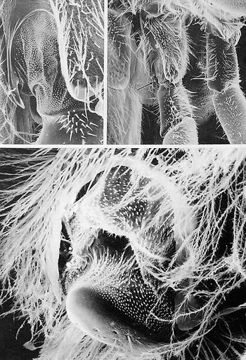
DRONELEGBASES
TOPRIGHT.Basesofthemiddle(mesothoracic)andhind(metathoracic)legs(theabdomenistotheleft).Intheforeground,proximallytodistally,arethecoxa,trochanter,andasmallportionofthefemurofthehindleg.Thearrowindicatesthetrochanter-coxaJoint,whichisfurthermagnifiedinthebottomrightmicrograph.(x60)
TOPLEFT.Scale-studdedintersegmentalmembrane(center)coveringthearticulationbetweenthehindcoxaandtrochanter.Theproximalsurfaceofthetrochanterhasabouttwodozenshort,sharp,socketedhairsthatmaybemechanoreceptorsthatcontactthecoxaInthecourseoflocomotionandinformthenervoussystemastolegposition.(x240)
BOTTOM.Trochanter-coxajoint(arrowintoprightmicropraph(x298)
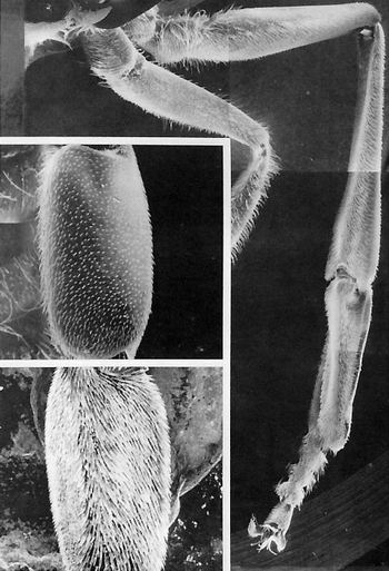
DRONEHINDLEG
TOP.Photomontageshowingentirelefthind(metathoracic)legandthecoxa,trochanter,andfemuroftheadjacentleg.Atthedistalextremityofthecompletelyexposedlegarethetwoclawsofthepretarsalsegment,followedproximallybyfourtarsalsubsegments,themostproximalofwhichconnectstotheelongateflattenedbasitarsus.Thebasitarsusarticulateswiththetibia.Thefemur,trochanter,andcoxaareinadistaltoproximalsuccession.Nopollengatheringortransfermodificationssuchasonthesesegmentsin'theworkerarepresentonthedronehindleg.ComparewithPlates2.28,2.29,2.30,and2.31.(x3l)
MIDDLELEFT.Lateralviewofthemetathoracicpretarsus(distaledgeatthetop).Thesetaearerelativelysparseandextremelyshort.(x37)
BOTTOMLEFT.Medialsurfaceofthemetathoracicpretarsus(distaledgeatthetop).Thissideischaracterizedbyadensepileofratherlongsetae.(x34)
DRONEHINDFOOT
BOTTOM.Pretarsusandfifthtarsalsegment.Theclawsgapewidelytobetterrevealthesoft,pursedmediallobe(arolium).Fivelong,curvedsetaeemanatefromthemediansclerite,which,inturn,articulateswiththelasttarsalsegment.Rowsofshorttricholdsensilladescendoverthedorsumofthelasttarsalsegment.(x180)
TOPLEFT.Threetarsalsegmentsofthehind(metathoracic)foot.Theplanta(seenhereastheuppersurface,whichisthebestorientationfortheviewer),coveredwithshorthairs,contactsthesubstratum.(x110)
TOPRIGHT.Lateralviewofthepretarsusandlast(fifth)tarsalsegmentshowingthecontractednatureofthearoliumandtheuprightorientationofthemedialsclerite.Thefivetrichoidsensillaarisefromthemedialscleriteatdifferentlevels.(x143)
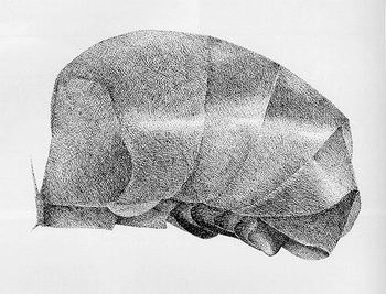
Lateralviewofthedronegaster(abdomen)
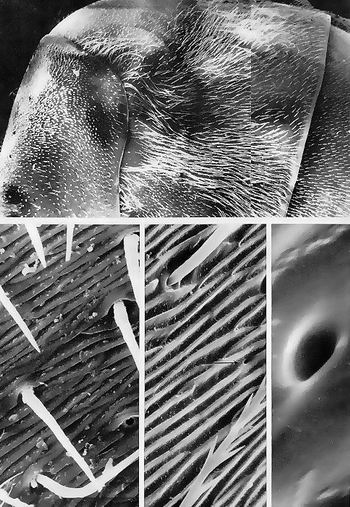
DRONEPETIOLE,DORSALVIEW
TOP.Slightlycurvedanteriormarginofthepropodeum,atthetopofthefield.Themembranousconnectiveofthe(dorsal)petioleisinthecenter.(x100)
MIDDLELEFT.Close-upofthedorsalmembranousregioninthetopmicrograph,withtheconnectivecut.Longitudinalwrinklesandcopiousscalesareprominentinthisregion.(x168)
MIDDLERIGHT.Apodemeforwingmuscleattachment,immediatelyanteriortothepetiole.(x234)
BOTTOM.Highermagnificationofthecuticularscalesofthedorsalmembranouswallofthepetiole.Fivetosevenspinesofseverallengthsprojectfromeachscale.(x3,230)
DRONEPETIOLE,ABDOMENDETACHED
TOPLEFT.Cross-sectionalcutthroughthemembranouspetiole.Thelongitudinallyseamed,scalymembrane(at12o'clock)andthethoraciccavityimmediatelybeneathareexposed.Twosizablemuscleswithsheaths(at3and9o'clock)arevisible;theseassistinelevatingtheabdomen.(x50)
TOPRIGHT.Reverseviewofthetopleftmicrographshowingthecrosssectionedanteriorendoftheabdomen(gaster).Thearrowpointstothehairplatethatisfurthermagnifiedinthemiddlerightmicrograph.(x50)
MIDDLERIGHT.Dorsolateralhairplateofthepetiole(arrowintoprightmicrograph).Thesemechanoreceptorsmonitorthegravitationalpullofthegasterontothethoraxviathepetiole.Thesocketed,unbranchedhairsaremechanosensors.(x300)
BOTTOMRIGHT.Highermagnificationofthesetaeofthehairplate.(X1,000)
BOTTOMLEFT.Longitudinalsectionthroughacuticularspurinthepetiolarregion.Thelaminatedappearanceoftheexocuticleisapparent.(x8,640)
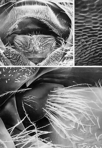
DRONEPETIOLARHAIRPLATE,OBLIQUEVIEW
BOTTOM.Petiole,thejunctionbetweenthefirstandsecondabdominalsegments.Thisjointensuresgreatflexibilityoftheabdomenrelativetothethorax.Monitoringthismovementandprovidingthebeewithinformationonthealignmentofthethoraxtotheabdomenrelativetogravityareseveral(mechanoreceptor)hairplatesthatareexternallyplacedproprioceptors.Onesuchhairplateisvisible;thesetae(sensilla)probablymakecontactwiththedorsalwallofthepetioleofthefirstabdominalsegment.Theblackandwhitediagonalbarindicatesthealignmentofbodysegments(theheadistotheupperleft).(x85)
TOP.Highermagnificationofthecontactzonebetweenthehairplatereceptorsandthedorsalpetiole.Thesefourdozensensoryhairsarearrayedinvariousdirections,soseveralofthemcontactthepetiolarprotuberanceatanyonetimeatanydegreeofflexion,whichensuresaconstantmonitoringofabdominalposition.(x430)
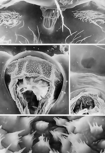
DRONESPIRACLES
TOP.Photomontageshowingprimarilythefirst(propodeal)andsecondsegmentsoftheabdomen(dorsumatthetop).Spiraclesarevisible(arrows)onthesetwosegments.Thewingshavebeenremoved.(x31)
MIDDLELEFT.Spiracleonthefirstabdominalsegmentatmoderatemagnification.Thisspiracleissomewhatlargerthantheothers,anditselongate-ovalrimdiffersfromthemoreroundedappearanceofthesucceedingspiracles.(x63)
BOTTOMLEFT.Close-upofthefirstabdominalspiracle.Theoperculum(plate)coveringthisspiraclehasbeendamagedinspecimenpreparation;intheintactbeethisplatecanbepulleddownbyanocclusormuscle,muchlikeawindowshade,thusshuttingoffthisspiraclefromtheoutside.Theoperculumisnevercompletelyopenanditsexcursionisnotlarge,sothissituationpermitsonlyaminimalaperture.(x170)
MIDDLERIGHT.Twospiracles,oneeachonthesecond(left)andthird(right)abdominaltergites.Themorecircularnatureoftheseopeningsisevident.Thesespiraclesarenoticeablylargerthanthoseoftheworkerandqueen.(x31)
BOTTOMRIGHT.Highermagnificationofthethirdabdominalspiracle.Thecirculardepressioninthecuticleleadstoanovalpit(lowerright)thatconnectsdirectlytotheunderlyingtracheaviaavalvemechanismcapableofoccludingthespiracleatriumfromthetrachea.Thebranchedhairsandvalvularapparatusmaybebarriersthatkeepmitesfromenteringthetracheathroughthisspiracle.(x250)

DRONESPIRACLES,CLOSE-UP
TOP.Surveyofaportionofthesecondandthirdabdominaltergitesshowingthespiracleonthethird.Theposteriormarginofthesecondabdominaltergite,whichissparselycoveredwithbranchedbodyhairs,isvisibleontheleft.Ontheanteriormarginofthethirdtergitearefewsetae,buttheteardrop-shapedspiracularopeningissurroundedbysocketedhairsonitsdorsal,ventral,andposteriormargins.(x175)
BOTTOMLEFT.Close-upofthespiracularopening.Insidethespiracleisthecuticularatrialwall,whichbulgesoutsomewhatandiscoveredwithveryshorthairs.Atmosphericairpassesoverthesefinersetaeandgainsentrancetothetracheadirectlybehindtheatriumviaasmallchannel.Thiscanalcanbeopenedwhenthemusclesofitsvalverelax.(x400)
BOTTOMRIGHT.Highermagnificationofthesmall,unsocketedhairsofthemedialwallofthespiracularatrium.Thefunctionofthesehairsmaybetoremoveparticulatematterfromtheairstream.(x800)
DRONEABDOMEN,DORSALSURFACE
TOP.Photomontageofthedorsalportionoftheleftabdomen.Abouthalfofthesecond,third,andfourthabdominaltergitesarevisibleintheirtotallateralexpanse.(x40)
BOTTOMLEFT.Highermagnificationofasectorofthelateralsurfaceofanabdominaltergiteshowingthefurrowlikecuticularrelief.Sparse,unbranched,socketedhairsarevisible.Thepores(at4and10o'clock)areprobablyorificesthroughwhichhairsonceprojected.(x775)
BOTTOMMIDDLE.Highlymagnifiedareaofabdominaltergiteshowingbothsocketedsmoothsetaeandabranchedbodyhair.Aporeisvisible(arrow).(x1,400)
BOTTOMRIGHT.Highermagnificationofacuticularporeonanabdominaltergite(arrowinbottommiddlemicrograph).Thisorificehasfoundedlipsandnocuticularlid.(x14,000
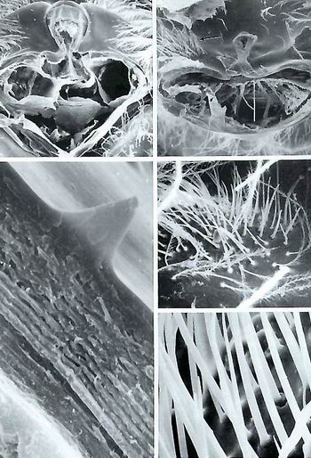
DRONEGENITALORIFICE
TOPLEFT.Terminalsegmentsoftheabdomen,frontalview.Two(paired)penisvalvesclaspthephallotreme,inwhichthepenisisbarelyvisibleinitsinvertedposition.Lateraltoeachofthepenisvalvesisa"bewhiskered"paramere.Dorsalandventraltothepenisvalvesaresectorsofintersegmentalmembrane.Withinthedorsalintersegmentalmembrane(left),justabovetheleftparamere,isthegreatlyreducedninthtergite.Thesclerotized"roof"fortheseorgansiscalledtheeighthtergite;immediatelybelowtheapexofthatroofisablackcrescent,theanus.Justbelowtheanusisaplatelikeareacalledtheproctiger.TheeighthsterniteformstheventralV-shapedenclosurefortheseterminalia.(x40)
TOPRIGHT.Cuticularsculpturingoftheintersegmentalmembranesurroundingtheparamere-penisvalvecomplex.(x484)
BOTTOM.Highermagnificationoftheleftparamereinthetopleftmicrographshowingkindsofsetaethatariseontheparameralplateandlieovertheleftpenisvalve.Ninesetaealsoarisefromthelateralportionoftheparamere.Immediatelyaboveandtotheleftoftheparamereistheverysmallninthtergite.Theeighthtergiteandsterniteformtheupperandlowerdiagonalscleritemarginsrespectively.(x168)
DRONEGENITALORIFICE,INTERIORVIEW
BOTTOMRIGHT.Frontalviewoftheterminalsegmentsoftheabdomen.Becauseofcollapseofthelastsegments,theproctigerandanusarehiddenundertheeighthtergite,whosearchedposteriormarginisvisibleatthetopofthefield.Thearrowpointstothephallotreme,whichisfurthermagnifiedinthebottomleftmicrograph.(x75)
BOTTOMLEFT.Phallotreme,theexternalopeningofadeependophallicpouch(arrowinbottomrightmicrograph).(x510)
TOP.Cuticularscalesonthedorsalaspectofthelast(seventh)tergite.Eachscalehasfourtosixspinelikeprocessesinessentiallytwosizeclasses.(x1,085)

DRONEGENITALIA
TOPLEFT.Vestibulum(bottomofthefield).Oneithersideofthevestibulumarethelateralhornsorcornuti(arrows).Theprincipalfeatureisthecervixofthetubelikepenis.Chevronlikescleritescovertheventralwallofthecervix.(x31)
MIDDLELEFT.Baseofthegenitalia,locatedimmediatelybelowthefieldinthetopleftmicrograph.Theventralplateofthevestibulumofthepenisiscoveredwithflexuoussetae.Onbothsidesofthevestibulumarethecornuti(externalgenitallobes).(x31)
BOTTOM.Tuftsofspiculesonthemedialwallofthecervixofthepenis.(x441)
TOPRIGHT.Highermagnificationofthewallsurfaceofthemalegenitalduct.(x300)
MIDDLERIGHT.Surfaceofthehorn(cornutus).Thedorsalandlateralsurfacesarepapillatewhiletheventralsurfaceisrelativelysmoothwithlongitudinalfolds.(x100)
SPERM
TOPLEFT.Floorofthecervixofthepenis.Profusesmallspines(spicules)linethisintima.Severalslenderspermarebarelyvisiblelyingatopthespicules.(x241)
BOTTOMLEFT.Massed,intertwinedspermatozoa.(x3,900)
TOPRIGHT.Oneentiresperm.(x700)
UPPERMIDDLERIGHT.Onespermhead.Thesmallnodulesoftheheadareprobablyamanifestationoftheacrosome.(x16,000)湖北天马养蜂场,加我们的微信一起学养蜂。
LOWERMIDDLERIGHT.Head(right)andtail(left)ofasperm.(x3,000)
BOTTOMRIGHT.Highermagnificationofthespermtail.Thelineargroovesmayrelatetotheinterspacesbetweenadjacentperipheralmicrotubulesingletsintheaxonemeofthetail.(x10,000)