这篇文章主要是从各个角度用显微镜观察研究蜂王,每个图片代表蜂王的一部分,图片下面都有相关信息的描述。
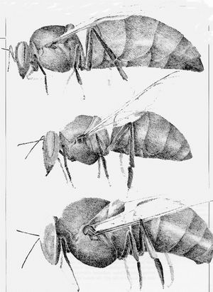
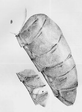
TOPTOBOTTOM:queen,workeranddronehoneybees.
drawnapproximatlytoscale.Bodyhairsomitted.(X8).
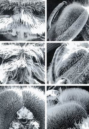
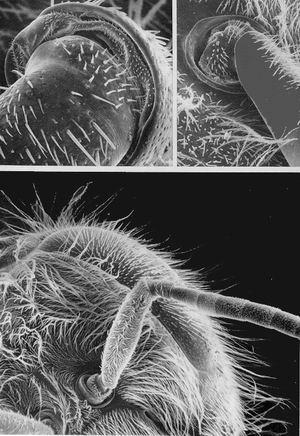
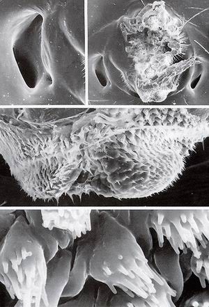
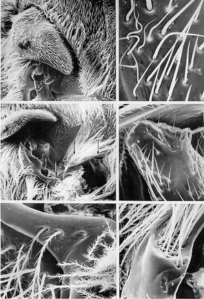
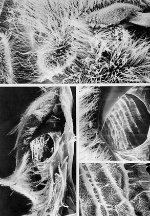
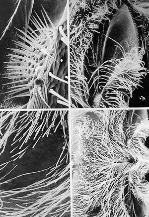
EYES,HONEYBEECASTES
TOPRIGHT.Workercompoundeye,lateralview.Theeyeissurroundedbytheshort-hairedgena(cheek).Thedense,coarse,longinterfacetalhairsoftheeyesareapparent.(x31)
MIDDLERIGHT.Queencompoundeye,lateralview.Comparewiththetoprightmicrograph.(x31)
BOTTOMRIGHT.Dronecompoundeye,lateralview.Thisandtheotherviewsoftheeyesshowasizablevisualfield,whichisgreatestinthedrone.Thedronebeecanseeforward,totheside,downward,upward,andtosomeextentrearward.(x31)
TOPLEFT.Worker,vertexofhead.Theantennaearevisible(bottomofthefield)butmostoftheotherstructuresareobscuredbyhairs.Intheuppercenter,thecircularoutlinesofthetwolateralocelli(butnotthemedianocellus)arebarelyvisible.(x31)
MIDDLELEFT.Queen,vertexofhead.Comparewiththetopleftmicrograph.Theocelliareobscuredbyhairs.(x31)
BOTTOMLEFT.Drone,vertexofhead.Thetworoundedeminences(topofthefield)arethedorsalsectorsofthecompoundeyes,whicharecontiguousinthisregion.Inthelowercenterofthefieldarethethreeocelli(oneisalmostcompletelycoveredwithhairs).Thesearelocateddownonthefaceofthedroneratherthaninthedorsoanteriorportionoftheheadasinthequeenandworker.Thedorsalbondingofthecompoundeyeshasapparentlydisplacedtheocelli.(x31)
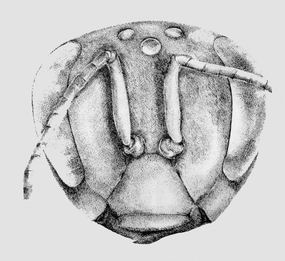
QUEENHEAD
FrontalViewofthequeenhead
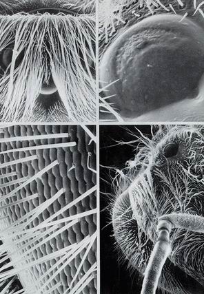
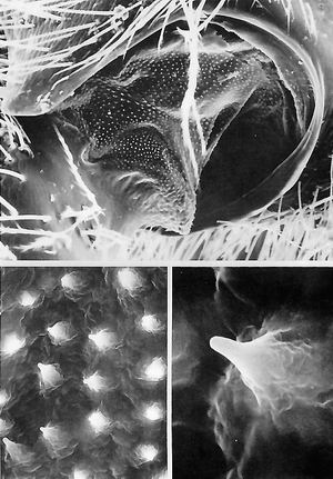
QUEENEYES
TOPLEFT.Trioofqueenocelli,twolateralandonemedian.Longbodyhairsdrapeoverthesesocalledsimpleeyes.(x58)
TOPRIGHT.Highermagnificationofthemedianocellus.Asinglelensservesasthedioptricapparatusforhundredsofunderlyingphotoreceptorcells.Theopticsaresuchthatnoimageisformedatthelevelofthephotoreceptorcells.(x600)
BOTTOMRIGHT.Surveymicrographofthequeencompoundeyeshowingitshirsutecharacterandtheglabrousmedianocellus(topofthefield).Thelateralocelliareobscuredbytheangleofviewandhairs.(x50)
BOTTOMLEFT.Interfacetalhairs,whicharesocketed,relativelylarge,andlong.Thedepthofthefocalplaneisevident,asthefocusismaintainedovertheconsiderablecurvedexpanseoftheeye.(x496)
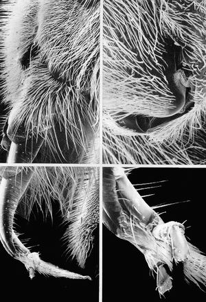
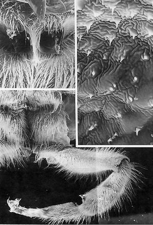
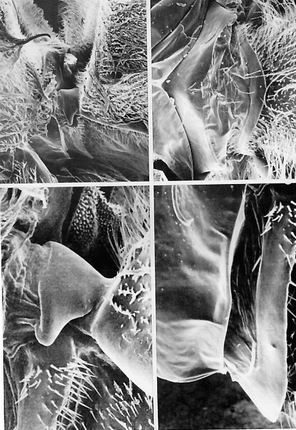
QUEENANTENNA,BASEANDELBOW
BOTTOM.Surveyfromthevertexofthequeenheadshowingthehairyhemisphericalcompoundeyewithitsarrayofinterommatidialhairs.Bothantennalbasesarepresent;theleftscape(firstantennasegment)istheprominentshortantennasegment,followedbythemuchlongerpedicel(thefirstsegmentafterthescape),andfiveflagellumsegmentsfollowthereafter,whilesixadditionalsegmentsareoutsidethisfield.Theflagellummovesrelativetothescapebytheactionoftwomusclesthatspanthepedicelandinsertonthefirstflagellumsegment.(x53)
TOPRIGHT.Baseofthescape,seatedwithinamembrane-linedsocketthatliesonthewallofthefrons.Presumedmechanoreceptivehairsareinrowsonthewallofthescape.(x110)
TOPLEFT.Close-upofthearticulationofthescapewiththeantennasocket.Astheantennamoves,thehairsonthescapebasemaybedifferentiallybentagainstthesocketside.Thesehairsmaybemechanoreceptorsthatmonitortheattitudeofthescape.(x330)
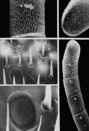
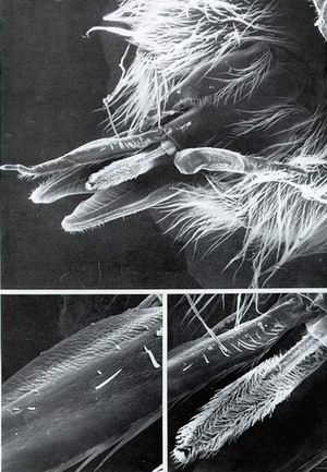
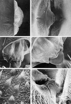
QUEENANTENNASURFACE
BOTTOMRIGHT.Lastfivesegmentsofthequeenantenna.Theantennasurfaceisprofuselycoveredwithavarietyofuniformlydistributedsenseorgans.Oneachsegment,usuallyattheproximalanddistalborder,aresmallclustersofpitorgans,whichappearasbrightspots.(x144)
TOPRIGHT.Terminalsegmentofqueenantenna.Afewstoutpegorgans(sensillabasiconica)andstoutlongpegsareseenamongthemajorityofsensoryhairs.Theplateorgansarenotvisible.(x270)
TOPLEFT.Medialsurfaceofthesecondantennasegmentofthequeen.Afewpitorgansshowbrightly.Itisnotknownwhythesepitorgansreflectmoresecondaryelectronstothescanningelectronmicroscopecollectorandthereforeappearbrighter.Thefaintcircularoutlinesoftheplateorgansarevisible.(x330)
MIDDLELEFT.Highermagnificationofthesurfaceonthepenultimatesegmentofthequeenantenna.Thebrightpitorgansarerelativelysparsebutareaggregated.Placoid(plate)andtrichoid(hair)sensillasurroundthepitorgans.(x2,200)
BOTTOMLEFT.Close-upofaplateorganandseveraltrichoidsensilla.Thecrackthatextendsalongtheouterrimofthisplateorganisprobablyartifactual.Atthismagnificationthesubtlecuticularribbingreliefaroundtheorgan'speripheryisvisible.Exquisitelysmallpores(notvisible)linetheseribsandpermitodorantmoleculestoenterandmakecontactwiththesensorydendritescircumferentiallyarrayedundertheplate.(x5,500)
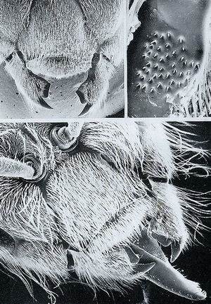
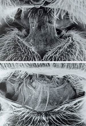
QUEENHEAD,ANTERIORVIEW
BOTTOM.Queenhead.Atthebaseoftheantennaeandprojectingdownwardistheroughlyrectangularclypeus;extendinginfrontofthatisthesmaller,largelyrectangularlabrum(upperlip).Bothmouthpartsareveryhirsute.Alongthelateralsidesoftheclypeus-labrumaretheclawlikemandibles.Projectingforwardfrombetweenthemandiblesarethegaleaandbehindthesearethelabiallobes.(x56)
TOPLEFT.Queenmouthparts,dorsoanteriorview.Herethemandiblesarebetteroutlinedandpartoftheirarticulationwiththegenaeisvisible.Atthisviewinganglethelabrumcompletelycoversthegaleaandlabiallobes.(x41)
TOPRIGHT.Medioproximalsurfaceofthemandiblewhereitadjoinstheclypeus.Over40small,socketedhairs(possiblymechanoreceptors)makecontactwiththesidesoftheclypeusinnormalmastication.Ifthesehairsareinnervatedandmechanosensoryinnature,theycouldmonitortheattitudeofthemandiblerelativetothatoftheclypeus-labrum.(x438)
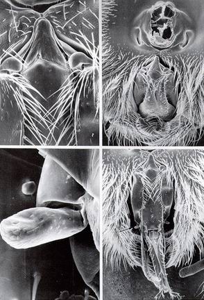
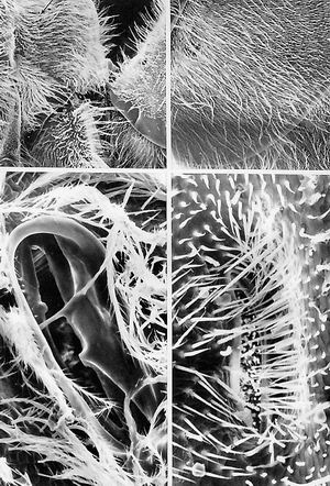
QUEENTENTORIALPITS
TOP.Frontalviewofantennae(top)andtheclypeuswithlabrum(bottom).Twoanteriortentorialpits(arrows),lyingontheepistomalsulcus,arecannular(tubular)structuresthatextendinwardtoformpartofthetentorium(internalskeleton)ofthehead.Thelatterprovidesgeneralstructuralsupportandcuticularbracesformuscleattachment.(x90)
BOTTOMRIGHT.Close-upoftherightanteriortentorialpit.Thisisoneoftheopeningsofthetwohollowcuticulartubesthatextendthroughtotheposteriortentorialpits.(x600)
BOTTOMLEFT.Highermagnificationofthequeenrightanteriortentorialpit.(x750)
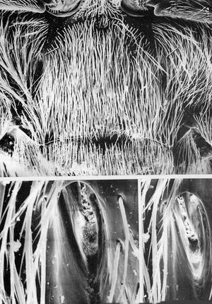
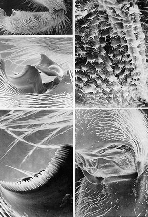
QUEENPROBOSCIS,LATERALVIEW
BOTTOMLEFT.Sharplybentglossa(tongue).Overhalfthelengthoftheglossa,theprincipalfeatureofthismicrograph,iscoveredbytheembracingproximalsegmentsofthelabialpalps.Thelabialpalpsterminateinapairofbudlikeappendagesthatappeartobesensoryinnature.Themostextendedappendageistheglossa.(x65)
BOTTOMRIGHT.Close-upoftheterminusofthelabialpalp.Each"bud"hastwosegmentsandbearsshortcuticularspinesandpegs,whichmaybesensorsfortasteandtouch.(x180)
TOPLEFT.Proximalportionoftheglossaandlabialpalps.Themandibleswiththeirnakedtipsincompletelycovertheglossaandthehairylabrumabutsandsomewhatoverhangstheparaglossae.(x65)
TOPRIGHT.Baseofthemandible(bottom)whereitarticulateswith(hingeson)thegena(top).Anarrayoftinycuticulardenticlesisvisibleontheintersegmental"membrane"(x169)
QUEENMOUTHPARTS,POSTERIORVIEW
TOP.Mouthparts,posteriorview.Theinnermostprojectingpiecerepresentstheretractedglossa(tongue),whichismuchsmallerthanthatoftheworker.Distallytheglossaistippedbya(brightappearing)flabellum.Atthebaseoftheglossaarethebilobedparaglossae;thesetwopiecesemanatefromaflattenedscleritecalledtheprementurn.Immediatelylateraltotheglossaarethetwofoursegmentedlabialpalps.Theoneontherightoftheglossahasbeenbentbacktobetterdisplaytheunderlyingmaxillarygalea.Whenfeeding,thetwopalpsandthetwogaleacometogethertoformatubeorfunnelthroughwhichliquidsaredrawnupbythemechanicalmovementsoftheglossaandthesuctioncreatedbythecibarium(notvisible).(x56)
BOTTOMRIGHT.Highermagnificationoftheundersideofthequeenglossashowingitsbilobedconstruction,flabellumatthetip,andthesparsehairs(sensillachaetica)ofthelateral-lyinglabialpalp.Afewveryshortpegs(sensillabasiconica)areseennearthesensillachaetica.Ofparticularinterestisthehighdensityandoverlapping(atthetips)natureofthesensillachaeticaontheglossa.(x121)
BOTTOMLEFT.Close-upoftheundersideoftheleftlabialpalpshowingthetwotypesofputativechemoreceptorsensilla,sensillachaetica(thelongerhairs)andsensillabasiconica(theveryshortbutstouthairs).(x327)
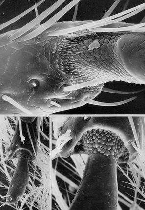
QUEENMOUTHPARTS,LABIALPALP
BOTTOMLEFTTWOdistalmostsegmentsofthelabialpalp.Lessthanadozensenseorgansareatthetip.(x388)
BOTTOMRIGHTTipofthepenultiniatelabialpalpsegmentandthebaseoftheapicalsegment.Chenioreceptorand/ormechanoreceptorhairsariseatthebaseofthedistalsegment.(x1,068)
TOPArticulationbetweenthefirstandsecondsegmentsofthequeenlabialpalp.Pegorgansandsensillachaeticaareobviousfeatures.Cuticularscales(probablyuninnervated)arecircumferentiallyarrayed.(x1,333)
QUEENHEADPOSTERIORVIEW
BOTTOMProboscisandothermouthpartsposteriorviewofthehead.Themouthpartsareextendedforward.Moststructuresintheothermicrographsofthisplatearevisibleinthisfield.Thearrowpointstothemaxillarypalp.(x40)
TOPRIGHTHead,posteriorview.Thelargeopening(lop)istheforamenmagnum,towhichisattachedthecervix(orneck,throughwhichpassthenervecord,aorta,andesophagusastheyspantheheadtothethorax).Theopposingcrescent-shapedaperturesflankingthebasalpartoftheforamenaretheposteriortentorialpits.Atthebaseoftheforamenisthetriangularpostmentum,whichissituatedatopthebilobedprementum.Embracingthelatterarethestipesandgalea.Herethemouthpartsareretractedbeneaththehead.(x37)
TOPLEFTHighermagnificationofthepostmentumregion.Flankingeachsideofthepostmentumisthecardo.(x112)
BOTTOMLEFTMaxillarypalp.Thislargelyunadornedstructureiswithoutobviouscuticularsensilla.Possiblythe"blebs"arecampaniformsensilla.(x564)
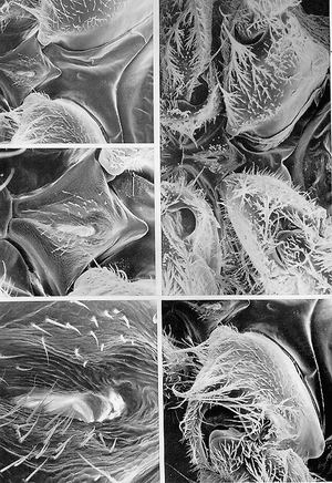
QUEENCERVIX,HEADDETACHED
TOPRIGHT.Occipitalforamenontheposteriorofthehead.Thisholecommunicateswiththecervix(neck),whichinturnattachestotheprothorax.Oneachsideoftheforamenaretheposteriortentorialpits,whichextendtotheposteriortentorialbridgewithinthehead.Betweenpitandforamenisthelateraloccipitalscleritewithitsrowsofmechanoreceptivehairs.(x66)
TOPLEFT.Close-upoftheleftposteriortentorialpit.Attheextremeuppermostedgeandbelownearthelowerrightedgeofthepitaresmallerdepressionsassociatedwiththedorsalandventralbridges.(x198)
MIDDLE.Ventralaspectofthebilobedcervicalmembrane,whichiscoveredwithshortcuticularspines.(x540)
BOTTOM.Close-upofthesecuticularspinesonthebilobedportionofthecervix.Upto20blunt,short,fingerlikeprocessesmayemanatefromeachscaleatitsdistalend.(x5,400)
QUEENCERVIX
TOPVentralcervix(neck)regionshowingtherathernarrowandmombranouscharacterofthecervix,whichconnectstheheadtotheroundedrightandleftepisternalplatesofthethorax(bottom)Thesurfaceofthecervixisstuddedwithcircumferentiallyarranged,tinycuticularscales.(x90)
BOTTOMDorsalcervixregion.Thecervix,withseveralfoldsandcoveredwithsmallcuticularstuds,visiblyconnectstheprescutumofthethorax(bottom)totheoccipitalregionofthehead.(x67)
QUEENCERVIX,CLOSE-UP
BOTTOMLEFTDorsoposteriorview(viewedobliquely)ofthepostocciputofthequeenwheretheheadjoinsthedarker-appearingmembranouscervix(neck).Longbodyhairscoverthethorax.Thearrowindicatesanareafurthermagnifiedinthetopleftmicrograph.(x84)
TOPLEFTLateraloccipitalsclerite(hairplate)(arrowinthebottomleftmicrograph)bearingputativemechanoreceptorhairsthatarebentwhenthisplatetouchestheepisternumoftheprothorax.Inbending,thesehairsinformthecentralnervoussystemoftheangleoftheheadrelativetotherestofthebody.(x912)
RIGHTHighermagnificationofthecuticularscalesadorningthemembranouscervix.Theadaptivevalueofthesescalesandtheirshort,pectinateprocessesisunknownatpresent.(x2,200)
QUEENTHORAX
Posteriorregionbearingtherelativelylargeovalspiracle(arrow)isactuallythefirstsegmentoftheabdomen(seealsoPlate1.36)althoughitisbroadlyfusedtothethorax.
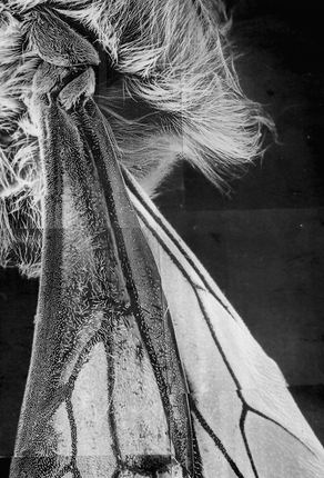
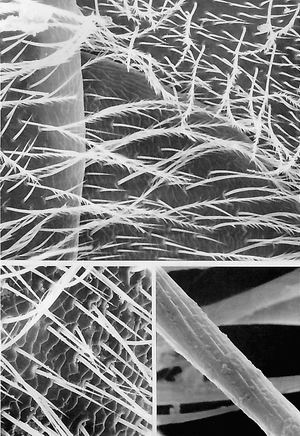
QUEENWINGS
Photomontageoftheproximalthirdoftheforewinganditsassociatedplatesatthepointofthewing'sarticulationwiththethorax(theheadIstothetop,dorsumtotheright).Visibleatthewinghingeareaisthelargestplate,thetegula,whichisclosesttothebody.Abovethetegulaandtotheleftisthehumeralplate;totherightisthemedianplate,whichseeminglycommunicateswiththevannalvein(thestoutveinthatprojectsapproximatelythroughthecenterofthewinginthisviewingangle).Thehighdensityandfinecharacterofthemicrotrichiacoveringthewingsurfaceisevident.Despiteitsratherstiffappearance,thewingisremarkablyflexibleandresilient,capableofpropellingthebeeatspeedsofover20kmperhour.Withtheirwings"disengaged"andfoldedbackoverthebody,queensareabletovibratetheirwingmusclestoproducethesoundsknownaspipingorquacking.(x34)
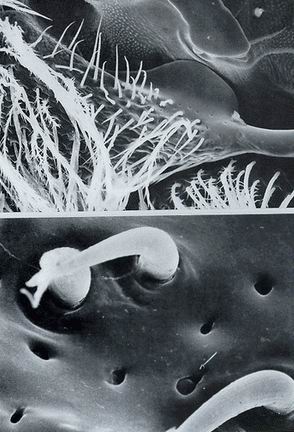
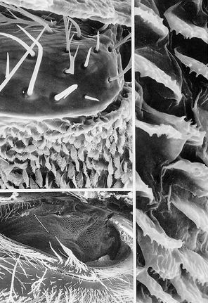
QUEENFOREWINGBASE
TOPForewingbaseshowingavarietyofhairtypes.Somefunctionalinferencescanbedrawn,basedonthesizeandpositionofthehairs.Shortandlongtrichoidsensillamayhaveanexoreceptivefunctionandrespond(bend)towindshearforces.Thelongonesmayalsobeproprioceptiveinnatureiftheycontactthepleuralsurfacewhenthewingisuprightandinformthecentralnervoussystemofwingattitude.Thebodyhairsareprobablynotinnervated.Chemosensorypitorgansarealsopresent.Behindthepits(lop)isanaxillaryscleritethatarticulateswithaportionofthepleuron.(x270)
BOTTOMHighermagnificationofthepitsandshorttrichoidsatthewingbase.Apitinthelowercenterofthefieldappearstohavearecesseddome(arrow)andmaybeacampaniformsensillum.Thehairandthepitmaybothhousesensoryneuronscapableofmonitoringthewingflexion.(x3,600)
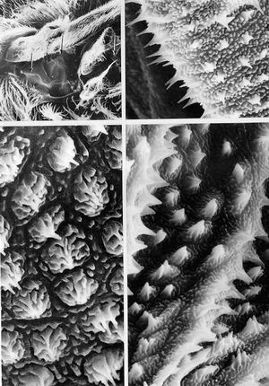
QUEENWINGBASEMEMBRANES
TOPLEFTForewingbaseandstubofthehindwing(centerrightof//I(,fieldInterestinglateralmembranousareasinthemiddleofthefield(arrows)areinhighermagnificationintheotherthreemicrographsofthisplate(seealsoPlates1.18,1.19,1.21,and1.22).(x55)
BOTTOMLEFTRosettelikecuticularspinesatthewingbasearrayedinrowswithhigh-relief,cuticularsculpturingbetweenthesespineclusters(leftarrowhitopleftmicrograph(x23,400)
TOPRIGHTPleuralsurfacebeneaththehindwingbase(rightarrowintopleftmicrograph).Acutelytippedspinesrisefromasurfacecoveredwithsmallcuticularknobs.(x2,200)
BOTTOMRIGHTPleuralmembraneatthebaseoftheforewing(centerarrowintopleftmicrograph)Elsewhereonthebodycuticularsculpturingischaracterizedbymoreprominent(thaninthetoprightmicrograph),short,stoutspinesinfurrowsbetweenrowsoftallerspines.Thecuticularsurfaceiscoveredwithaseriesoflow-lyingridges.(x23,800)
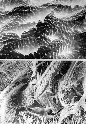
QUEENWINGARTICULATION
BOTTOM.Articulationatthequeenwingbase.Theleadingmarginalveinsoftheforewingandhindwingpivotfromaxillaryscleritesandprojectdiagonallyinthismicrograph.Aportionoftheventralwingmembrane(centerofthefield)possessesnohairsbutratherhaslow-lyingcuticularprotrusionsandmultipeakedridges(topmicrograph).(x95)
TOP.Forewingbase.Theundersideofthewingexhibitsseveraltypesofcuticularrugosites.Thefunctionofthissurfacereliefisnotknown.(x5,100)
QUEENWINGBASESCLEROTIZEDPLATES
TOPLEFTBaseoftheforewing(theheadistothetipperleft).Thehairytegulapartlycoversthetriangularhumeralplate.
TOPRIGHT.Hairplateofsocketed(presumed)mechanoreceptorsontheinterioredgeofthehumeralplate.Electronbeamdamageprobablycausedthebendingandtwistingofhairs.Rotationalmovementsofthetegulaandhurneralplatebendthesehairs;ifinnervated,theycouldmonitorsuchdislocations.(x66)
MIDDLELEFTAnotherviewoftheforewingbaseshowingtheupraisedhumeralplate.Thearrowspointtostructuresshowninfarhighermagnificationcationonthebottomleft,bottomright,andmiddlerightmicrographs(X1'50)
BOTTOMClose-tipoftheradiusvein.Branchedhairsemanatefromthispartofthewingbase.Structuressurroundingthisveinbasearevisibleinthemiddleleftmicrograph,indicatedbytheleftarrow.(x2,600)MIDDLERIGHT.Condylelikeprojectionatthewingbasewithhairsaparentlylyincontactwiththewingmargin.Hairsprojectatdifferentlevelsandanglesfromthisknob.Thesehairs,ifinnervated,maybemechanoreceptorsrecordingwingbeatfrequencyand/ortensionandflexionofthewings.Alow-magnificationviewofthisstructureinrelationtootherwingstructuresisinthemiddleleftmicrograph(rightarrow)(x300)
BOTTOMRIGHTHigh-magnificationviewofafascicleofabout20socketedhairsinacuticularprotuberanceatthewingbase.Featuresadjacenttothisstructurearevisibleinthemiddleleftmicrograph(middlearrow)Thesehairs,ifinnervated,maybemechanoreceptorsmonitoringasinglekindoftemporaldisplacement,suchastheupstrokeordownstrokeofthewing.(x60)
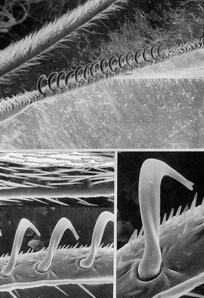
QUEENW1NGHOOKS
TOP.Hooksonthehindwingclosetothethickenedbaseofthearticulatingforewing.Particularlyapparentarethehind-winghairsontheleadingedgeofthehindwing(comparewithpegorgansoftheworker,Plate2.23).Thesehairsprojectforwardandarefoundoneithersideofthe15hooks.(x170)
MIDDLELEFT.Highermagnificationofthetrailingedgeoftheforewingclaspedbythewinghooksinflight.Thehairsadjacenttothetrailingveinoftheforewingaresocketedandmaybemechanoreceptorscapableofsensingtheattackangleofthewings.Thebenttipsofafewhairsmayhavebeencausedbyelectronbeamdamage.(x480)
BOTTOMLEFT.Hooksontheleadingedgeofthehindwing,eachwithaslightlyforkedterminus.Thebentandtwistednatureofthehookisapparentfromthisangle;inthreedimensionsthehookextendsintwodirections.(x605)
BOTTOMRIGHT.Enlargementofthemiddlehookinthebottomleftmicrograph.Thesocketforthehookislesspronouncedthanthatoftheworkerandnopegorgansarepresent,onlysocketedmicrotrichiathatmaybecapableofmonitoringtheproximityofthewings.(x1,210)
QUEENFOREWINGSURFACE
TOPLEFTDorsalsurfaceoftheforewingshowingadensepopulationofshort,slender,socketedhairsthatmaybemechanoreceptorsthatprovideaerodynamicsense,thatis,monitorthevectorandmagnitudeofaircurrentsoverthewingsurface.(x550)
TOPRIGHT.Highermagnificationofasinglehair,revealinganovalratherthanroundsocket.Presumablythehair'sflexionisconfinedtothelongaxisoftheovalsoitcanrecordwindinoneaxisonly.(x3,300)
MIDDLE.Ventralsurfaceoftheforewing.Thesetaearelessdensethanonthedorsalsurface.Ifinnervated,someunsocketedhairsmaybechemoreceptors.(x510)
BOTTOMLEFT.Threeunsocketedsetaeontheventralsideoftheforewing.(x1,100)
BOTTOMRIGHT.Socketedsetaontheventralsideoftheforewingvein.Thetipcannotberesolvedwell,butitsbulbousnaturesuggestsaterminalpore.Ifso,thishairisprobablyachemoreceptor.(x5,500
QUEENHINDWINGSURFACE
TOPLEFT.Dorsalsurfaceofthehindwing.Numeroushairsareevenlyspacedoverthesurface.Winghooksarevisibleontheleadingedgeofthewing.(x168)
TOPRIGHTClose-upofthewinginthetopleftmicrograph.Socketedhairsarisefromtheveinandnonsocketedhairsareinthemembranous,interveinalarea.Veinsareoftenconduitsfornerves,sothesocketedhairsmaybeinnervatedandhaveamechanoreceptorfunction.(x800)
BOTTOMLEFT.Ventralsurfaceofthehindwing.Thesparse,scattered,nonsocketedhairsgivetheappearanceofpegorgans(sensiIlabasiconica).(x450)
BOTTOMRIGHTHighermagnificationofahair,whichlookslikeagroovedpeg,ontheventralhindwing.Suchhairsmaybechemoreceptors.Thedepressionorpit(bottom)delimitsthebaseofahairontheoppositesideofthewing.(x4,200)
QUEENPROTHORACICSPIRACLE
Top.Prothoracicspiracle,whichliesbeneaththespiracularlobe,orhairyplate(arrow)(theheadistothetopleftofthisobliqueview).Theforewingbaseisvisible(upperright).Thisspiracleisprotectedbythespiracularlobeandshroudedwithbodyhairs,whichmustberemoved,asintheothermicrographsinthisplate,beforethespiracleanditsinteriorcanbeobserved.(x56)
BOTTOMLEFT.Portionofthespiracularlobe,theatriumofthisspiracle,anditsimmediateinterior.Thisareaisrevealedwhenthehairsareremovedfromthethoraxandasuperficialsliceofcuticleistakenfromthisregion.(x1,500)
MIDDLERIGHT.Close-upofthetrachealatriumrevealingtheairwaywithoutanyofitscuticularovercoats.Theinnerwalloftheatriumhascuticularridgesfromwhichprojectsmallspinesorhairs,whichsupporttheintegumental"pocket"conceptoftheatrium.Thetrachealopeninghasbeenremoved.(x300)
BOTTOMRIGHT.Highermagnificationofthecuticularhairsandridgesoftheatriumoftheprothoracicspiracle.Thecuticularreticulumextendsalongtheflooroftheatrium.(x1,200)
QUEENFORELEG
BOTTOM.Photomontageoftheprothoracicleg(foreleg).Thethreemainsegmentsoftheleg(distaltoproximal)arethetarsus,tibia,andthefemur(thelargestofthethree).Attheproximalendofthetarsus(basitarsus)isthesharplynotched"antennacleaner."Aclosinglobe(fibula)orspurextendstopartlycoverthenotch.Ingrooming,theantennaispassedrepeatedlythroughthisnotchtocleanthesensoryhairsandplatesoftheantenna.(x34)
TOPLEFTProthoraciclegbases(legsremoved;headistothetop).Thetwoemptycoxalcavitiesaresidebyside.Thetipsofthemaxillae(top)aresituatedbetweenthecoxalcavities.(x60)
TOPRIGHTHighermagnificationoftheintersegmentalmembranebetweenthetrochanterandfemur.Theirregularlyfurrowedcharacterofthissurfaceisinteresting,asaretheunsocketed,tinyteatlikecuticularspurs.(x2,400)
QUEENFORELEG,CLOSE-UP
TOPLEFT.Surveyofthebasalportionoftheleg.Thesegments(fromlefttoright)arethebasitarsus,tibia,and(projectingata45'angletothetibia)thefemur.The"beak"attheendofthetibiaisthefibula.(x31)
MIDDLELEFT.Basitarsus(left),whicharticulateswiththetibia.Thelobeonthetibiaisthefibula,whichclosesoverthehairednotchforantennacleaning.(x120)
BOTTOMLEFTAntennacleaner,orcomb.Thissemicircularfringeofstiffhairsislocatedonthebasitarsusatitsjunctionwiththetibia.Theantennaispulledpastthesehairstoremovedebris.Whentheforelegisflexed,thenotchwithhairsisoverlaidbytheshortfibula,formingacuticularcirclethatenablesallsidesoftheantennatobecleanedatonce.(x384)
BOTTOMRIGHTPosteriorportionofthefemur-tiblaJointoftheforeleg.(x144)close-upofthecuticularreliefandtheshortspursthatadornthebackofthefemur-tibiajoint(intersegmentalmembrane).Acomparisonofthisfieldwiththecorrespondingareainthebottomrightmicrographshowsthelimitedareaoverwhichthiskindofcuticularornamentationoccurs.(x168)
QUEENMIDDLEANDHINDLEGBASES
TOPRIGHTVentralviewofthemiddleandhind(rigt)legbases.Thecoxaeandtrochantersofthebind(metathoracic)legsareontheright,thoseofthemiddle(mesothoracic)legsontheleft.Therectangularplatebetweenbothsetsoflegsisthesternellum.Therhomboidalplateboundedbythefourlegbasesisthepropodealsternum;itispunctuatedbyacenterapodeme.(x47)
BOTTOMRIGHTVentralviewofthebaseofthemetahtoracicleg.Themajorappendagehereisthemetathoraciccoxa.Thedistalappendagethatprojectstothebottomofthefieldisthetrochanter.(x55)
TOPLEFT.Sternalscleritesituatedbetweenthemetathoraciccoxae.Thisplateismoreproperlycalledthepropodealsternum.(x50)
MIDDLELEFTHighermagnificationofthepropodealsternumandtheadjoiningcoxaeofthemesothoraciclegs.Thehairplates(arrow)atthebasesofthecoxaeareprobablymechanoreceptorsmonitoringlegmovement.(x100)
BOTTOMLEFTApodeme(cuticularinvagination)andthesurroundingpropodealsternum.(x300)
QUEENMIDDLELEGBASES
BOTTOMRIGHTThorax,ventralview.Themiddle(mesothoracic)coxaeareontheextremeright,oneabovetheother.Thegroove(line)runningsidetosidethroughthemiddleofthefieldisthemediansulcus.Thisdivisionseparatestherightmesosternumandmesoepisternumfromitsleftcounterpart.Thearrowpointstooneofthe"pincushion"structuresfurthermagnifiedinthetoprightandtopleftmicrographs.(x40)
TOPRIGHTHighermagnificationoftheleftcoxainthebottomrightmicrograph.The"eyelid"cuticularstructureisanintersegmentalmembrane,andattheextremebasalportionofthefieldisasmallportionofthecoxalcondyle.Immediatelyabovethecondyle(arrow)isthe"pincushion"structureindicatedbythearrowinthebottomrightmicrograph:thisstructureisevenfurthermagnifiedinthetopleftmicrograph.(x96)
TOPLEFTForty-oddtinysetae.Thismaybeacollectionofinechanoreceptors(makingupahairplate),whoseprobablefunctionistoirionitorlegloadingandpositionbyspinedisplacementthroughcoxalcontactwiththebasalmarginofthemesosternum(x664)
BOTTOMLEFTHighermagnificationofoneofthetwohairlessareasvisible(at7and11o'clock)inthesurveyoftheupperventerofthethoraxinthebottomrightmicrograph.Nocuticularhairsexistinthis(the7o'clock)areanorarethereanyemptysocketsorotherindicationsofcuticularsensilla.Alsoabsentisthenormalcuticularrelief.Thefunction,ifany,oftheseareasremainsamystery.(x208)
QUEENMIDDLELEGBASE
TOPClose-upofthelegbaseandcoxalcavityofthemiddleleg.Thearticulating(intersegmental)"membrane"ischaracteristicallycoveredwithrowsoftinyspinelikeprocesses.(x324)
BOTTOMLEFT.Close-upofthecuticularspinesarisingfromtheintersegmental"membrane"betweenthemesothoraciccoxaandtrochanterofthequeen.Thelinearityoftherowsofthesespinesisapparentinthisview.(x4,800)
BOTTOMRIGHT.Close-upofoneoftheunsocketedspinesshowingitsgeneralmorphologyandblunttip.(x18,000)
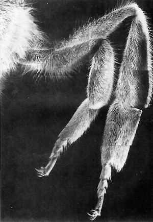
QUEENMIDDLEANDHINDLEGS
Photomontageofthemiddle,ormesothoracic(left),andhind,ormetathoracic(right)legs.Atthelegbasethefirstsegment,thecoxa,isalmostcompletelyobscuredbythehairsofthemesothoracicpleurites.Thenextsegment,thetrochanter,isvisible,extending(horizontally)toarticulatewiththelargerfemur.Thetibiajoinsthefemur(atthe"knee")andextendsdownward.Themesothoracictibiaisaboutaswideasthefemur,butthehindtibiaisflattenedandmuchbroader.Theseconddownward-projectingsegmentisthebasaltarsomere,whichisclearlymuchlargerthantheother,moredistaltarsalsegments.Inthequeenthemetathoracicbasaltarsomerelacksthepollencollectionapparatusofitscounterpartintheworker(seePlates2.28,2.29,2.30,and2.31).Fourremainingtarsalsegmentsarepresent;thelastone(pretarsus)iselongateandbearsclaws.Thesefirstfourtarsalsegmentshavenomuscles,butacommontendontraversesallofthemandinsertsintotheflexormuscleofthepretarsus.(x28)

QUEENMIDDLELEG
TOPLEFTArticulationbetweencoxaandtrochanterofthemiddleleg.(x140)
TOPRIGHTDigitiform,scale-coveredcuticularspuratthetibia-basitarsusjoint.(x31)
BOTTOMHighermagnificationofthetinyspurinthetoprightmicrograph.Itappearstoarisefromtheintersegmentalmembranebetweenthetwolegsegments.Thisminiscule,pineconelikespinemaybeanexternalproprioceptor-mechanoreceptorthatmonitorslegflexionortorque.Thecomparableappendageinworkerbeesiscalledthewaxspur(seePlate2.27).(x360)
QUEENFOREFOOTANDHINDFOOT
TOPPretarsusofthemetathoracic(hind)leg.Itstwomainfeaturesarethepairedclawssurroundingthetounglikelikearolium.Aboutahalfdozenlong,large,curvedhairsprojectbackwardfromthemediansclerite.Atthebaseofthearolium,andprojectingproximally(leftward),istheplanta,whichiscoveredwithspinesthatextenddistally.Theunguitractor,whichisnotwelldisplayedinthisfield,islocatedtotheleftoftheplanta.Themedialsurfaceofeachbilobedclawhasseveralranksoffinehairsandafewchaeticalikesensilla.(x270)Metathoracicpretarsusfacingtheviewer,dorsalsideup.
BOTTOMRIGTThemedianscleriteisvisible,alongwiththestoutspinesthatissuefromit.Severaldifferentformsofhairarepresentonthebaseofthelasttarsalsegment(top).(x168)
BOTTOMLEFTSpinyarolium.Astheclawsrelax,thearoliumassumesthepursedshape.Whentheclawsareactiveandextended,thearoliumisdrawnupwardandspreadsoutbetweentheclaws.Thebeeusesthisfleshylobetogripsurfacesthatcannotbepenetratedorgraspedbytheclaws.(x378)
QUEENABDOMEN
Gaster(abdomen),whichconsistsofafirst,ratherindistinctsegment(attachedtothethoracicregion)withaprominentspiracle,thepetiole(waist),andtheremainingposteriorgastersegments.Thedarkspotsindicatetherelativepositionofspiraclesonthefirstabdominalsegmentandonthatportionoftheabdomenbehindthepetiole.Thereisonespiracleoneachsideofthefirstsevensegments(seealsoPlate1.16).
QUEENI-PETIOLELATEPALANDVENTRALVIEWS
TOPLEFTLateralviewofthepetiolarregion,or"waist,"theconstrictedareabetweenthefirstandsecondabdominalsegments.Ontheleftisthe"bump"ofthepropedeum(firstabdominaltergalplate);ontheright,acrossthenarrowpetiolaristhmus,isthesecondabdominaltergite.The1,saddle"betweenthetwosegmentsisthemembranousroofofthepetiolepocket.(x61)
BOTTOMLEFTClose-upofthepetiole.Immediatelybelowandontheleftisthefirstabdominalsternite.Ontherightisthesecondabdominalsternite.(x210)
TOPRIGHTVentralviewofthepetiole.Amembranousareadividestheringlikefirstabdominalsternite(left)fromthecuticularringthatisthesecondabdominalsternite.(x55)
BOTTOMRIGHTPetiolarregion.Ontherightistheringlikesecondabdominalsternite.Projectingleftwardfromthatsterniteisthewrinkledandflexibleintersegmentalmembrane.(x175)
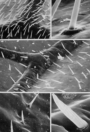
QUEENPETIOLE,DORSALVIEW
TOPLEFT.Dorsalviewoftheessentiallyhairlesspetiole.Thedarkverticalsector(withonesocketedcuticularhair)istheposteriorportionofthemesoscutellumTheintersegmentalmembranespanningthepropodeumandsecondabdominaltergiteappearsasatrilobedstructure.(x140)
MIDDLELEFT.Tangentialorientationofthepetiole(theheadistothetop).Thenumerouscuticularspinesintheinterseginentalmembraneareinasomewhatorderedarray.(x150)
BOTTOMLEFT.Highermagnificationofthemultipeakedcuticularspinesontheintersegmentalmembraneofthepetiolarregion.Theinterspineareashavealow-lyingbumpytexture.(x2,600)
TOPRIGHTPetiolarregion(theheadistotheleft).Thepropodeumhasbeentippedforwardsothatitsfullposteriorextentisvisibleasitattachestothegaster(abdomen).Theslenderpetiolarattachmentisthevitallinkagethatconnectsthesphericalthorax(withthefirstabdominalsegment)withtheevenmoremassivegaster.(x66)
MIDDLERIGHT.Dorsalviewoftheinterseginentalmembraneofthepetiolarregion(theheadistotheupperleft).Comparethismicrographwithitsleftsidecounterpartinthebottomrightmicrograph(x175)BOTTOMRIGHTLateralviewofthepetiolarregion.Attheextremelowerright(arrow)isthedorsalportionofthesecondabdominalspiracle.(x60)
QUEENABDOMINALSPIRACLES
TOPLEFT.Surveyofthepleuralportionofthefirstandsecondabdominalsegments.Thesmallspiracularapertures(onepersegment)(arrow)areobscuredbyextensivehaircover.(x31)
BOTTOMLEFT.Firstabdominalspiracleonthepropodealsegmentofthethorax.Thisisthelargestspiracle(alongwithitsoppositesidetwin).Itselevatedcuticularrimsurroundsashallowatrium,andtheinteriorrimisthetrueopeningintothetracheae.Thetracheaeareclosedofffromtheinner(interior)rimbyasizablevalve,whichisincompletelySeenherebecauseofathinfilmofdriedmaterialemanatingfromthetrachea.(x240)
TOPRIGHT.Abdominalspiracle(upperleft)Withdifficult\,,thecovertandlinearcharacterofthisaperturecanbemadeout.(x37)
BOTTOMRIGHT.Highermagnificationofaiiabdominalspiracle.Acuticularhairfenceoftergalorignincompletelycoversthisaperture,andseveralformsofhairsarepresent.TheinnerwalloftheatriumIsrepletewithveryshorthairs.(x360)
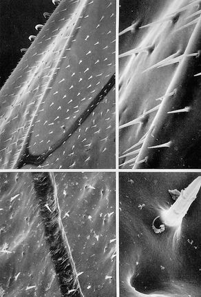
QUEENTHORAXANDABDOMEN,CUTICULARHAIRS
TOPPortionsoftwoterminalabdominalsegments.Ofinterestisthecuticularreliefthatextendsoverthesesegmentsandthefringedandglabrouscuticularhairsthatareabundantovertheentirebody.(x263)
BOTTOMLEFT.Cuticleandcuticularhairsofametathoracicpleurite.Mostofthesecuticularhairsareunfringedandunbranched,andthetipsaresharplypointed.Someofthelongbranchedhairsalongtheperipheryoftheabdominaltergitesandsternitesareattachedtoglandcellsthatmaybethesourceofqueenpheromoneobtainedbyworkersduringgrooming.(x550)
BOTTOMRIGHT.Close-upofacuticularhairshaftonanabdominalsternite.Theflutednatureofthesidewallisnotartifactual,althoughthe"blebs"ontheshaftwallmaybedebris.(x550)
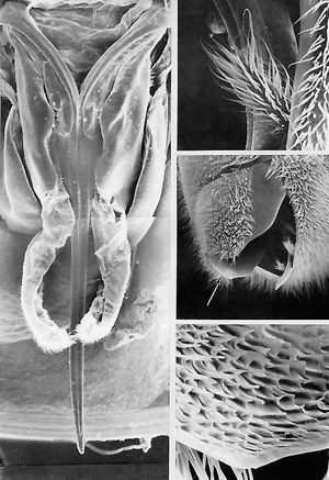
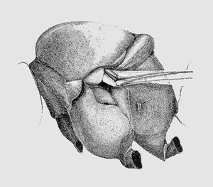
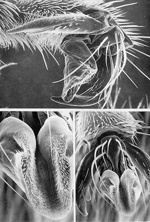
QUEENSTING
LEFTPhotomontageshowingtheentireventralsurfaceofthesting.Thetwobarbedlancets,incloseapposition,overlaythestylettroughtoformthevenomchannel.Oneithersidearethedistorted(inspecimenpreparation)sheathlobesofthesting.Eachsheathlobeisextendedproximallyfrombeneathanoblongplate.Medialtoeachplate,nearthetopofthemontageisaforkedstructure;each"tine"orramusconnectsproximallywithatriangularplate,whichinturnisassociatedwithaquadrateplate.Thelatterscleritetiesalongside(totheOutsideof)theoblongplate.SeetheAppendix,Fig.A.1.(x94)
MIDDLERIGHTLateralviewoftheabdominaltip.Theultimateabdominalsegmentseenhereisnumberseven;theseventhtergalandsternalplatesenclosemostofthestingaswellassegmentseighttoten.Theaperturethroughwhicheggsandfecespasswhenexpelledfromthebodyalsofunctionsastheentrancetothestingchamber.Thefuzzyprojectionsarisingfromthefloorofthestingchamberarethetipsofthestingsheath.Thearrowatthebottomleftofthefieldpointstotheareafurthermagnifiedinthebottomrightmicrograph.(x31)
BOTTOMRIGHTMedialsurfaceoftheabdominaltip(ventralsclerite)revealingmyraidsofstoutcuticularspines(arrowinrightmiddlemicrograph)(x62)
TOPRIGHTHighermagnificationoftheabdominaltipintherightmiddlemicrographshowingthesetoseconditionofthestingsheathatitsproximalorigin.(x250)
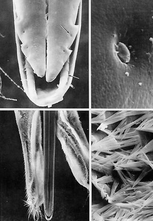
QUEENSTING.CLOSE-UP
TOPLEFTDistaltipofthestingapparatusshowingtheterminusofthevenomchannel.Twobarbedlancetsintheforegroundopposeeachother(earlierworkersreportedthatthequeenstinghadnobarbs).Behindthelancetsisthetroughlikestylet.Whenstinging,thelancetsslidebackandforthonserratedtracks(lowerarrow).Thetracksformthetroughatthetipofthestylet.Eachthrustpullsthestingdeeperintothevictim.Boththetrackandthegrooveineachlancet(notvisible)areserrated,probablytoreducefriction.Notethesmall"indentations"(oneperbarb)(upperarrow)onthelateralmarginsofeachlancet(seethetoprightmicrograph).Incomparisonwithworkerbarbs,thequeenbarbsarelessformidable,whichmaypermitthequeentostingrepeatedlywithoutlossofthesting.Otherworksuggeststhatthequeenstingiscoveredwithasurfacelubricantwhilethatoftheworkerisnot.(x1,040)
TOPRIGHTHighlymagnifiedviewoftheindentationindicatedbytheupperarrowinthetopleftmicrograph.Oneindentationisassociatedwitheachbarbonthelancetsandstylet.Thisstructureappearstobeacampaniformsensillum.Ifso,suchaproprioceptormightmonitorpressuregeneratedbytherelativedepthofinsertionofthestingorflexionofthelancets.(x15,000)
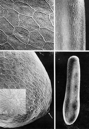
EGG
BOTTOMRIGHTEggproducedbythequeenbee.Bothendarerounded,andonepoleseemslargerthantheother.Atthismagnificationthecuticularreliefcharacteristicforthisspeciesisbarelyvisible,(x110)
TOPRIGHTHighermagnificationoftheeggsurfaceshowing,thereticulatedpatternofthecuticularridges.(x400)
BOTTOMLEFTClose-upofthelargepoleoftheeggshowingthelinearridgesystemenclosingpolygonalareasoftheeggsurface(chorion).Thearrowindicatestheareafurthermagnifiedintheinset.(x600)湖北天马养蜂场,加我们的微信一起学养蜂。
BOTTOMLEFTINSETMicropyleoftheegg.(x600)
TOPLEFTPolygonalzonesonchorion.Thebasalchorionsurfaceisrandomlystuddedwithminuscule,discretebumps.(x1,250)