这篇文章用显微镜从雄蜂各个角度进行拍摄说明,一张图片为一个角度,图片下面为相应的解说文字。
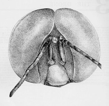
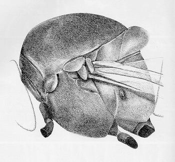
DRONEHEAD
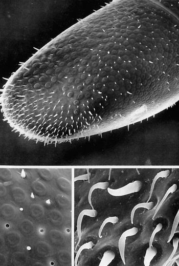
Frontalvieofthedronehead.
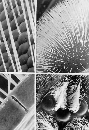
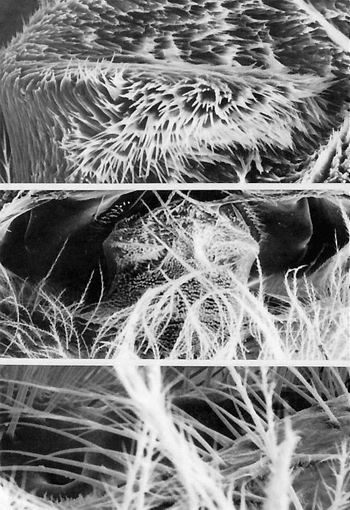
DRONECOMPOUNDANDSIMPLEEYE
TOPRIGHT.Compoundeyecoveredwithinterfacetalhairs.Theseinnervatedsetaeariseintheinterstitialspacebetweenadjacentfacetsofthecompoundeye.Atthebaseofeachhairisasinglebipolarneuron.Thesehairsaremechanoreceptors;bybendingtheymonitordirectionofwindcurrentsandairspeed.(x140)
TOPLEFT.Highermagnificationofthecornealsurfaceofthecompoundeyeshowingthesocketedbaseofeachhairandthehexagonaloutlineofeachlensfacet.Lensfacetsattheperipheryoftheeyeoftenhaveotherfrontalviewgeometricshapes.(x1,400)
BOTTOMLEFT.Longitudinalsectionthroughthecorneallensofthecompoundeye.Thedeepcleftthroughthelensmarkstheboundaryofoneommatidium;thisrecessalsoprovidesthespacenecessarytohousetheneuronandgliaofthehair.Thebiconvexdesignofeachlensfacetandthecuticularlaminationsarealsoevident.(x1,650)
BOTTOMRIGHT.Onemedialandtwolateralocelli.Thesethree"simple"photoreceptororgansarelocatedonthevertexoftheheadbetweenthetwocompoundeyes.Ocelliusuallydonotformimages.Thesephotoreceptorsmaymonitorlightlevelsandpossiblyprovideinputtosomephotoperiodicallyentrainedcircadianrhythms.Nofunctionisknownfortheprofusebundlesoffinehairsbetweentheocelli.(x60)
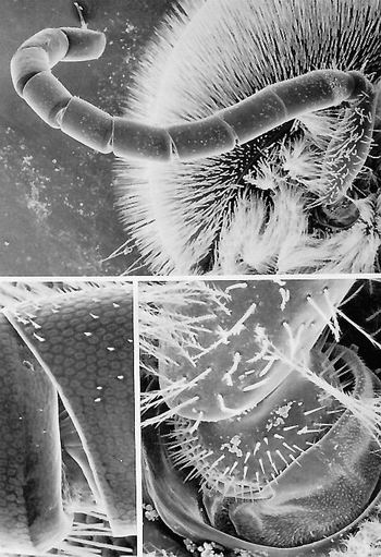
DRONEANTENNAANDBASE
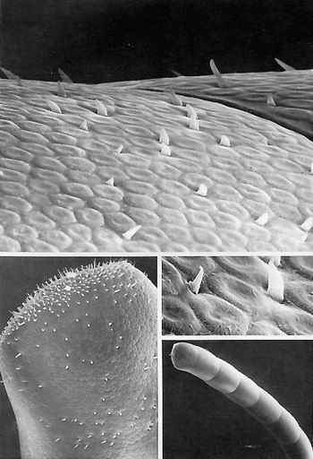
TOP.Droneantenna.Therearetwelvesectionsoftheantenna.Thedroneantennahasfarfewertrichoidsensilla(theveryshortwhite11whiskers"ontheflagellum)thandoqueenandworkerantennae(seePlates1.6and2.4).(x53)
BOTTOMRIGHTHighermagnificationoftheantennalbaseshowingthebasalknobofthescapeinsertingintothemembrane-linedantennalsocket.Microtrichiaareabundantoverthis"membrane."Ranksofsocketed(putative)mechanoreceptorhairscompletelyencirclethebase.(x347)
BOTTOMLEFTArticulationbetweentwoantennal(flagellar)sections.Eachsubdivision(theyarenottruesegments)fitsintothesucceedingonewithoutanyrealarticulationormusculature.Thebaseofeachannularportioninsertsintoasmallcavityoftheonebehindit.Spinesareabsentontheintersegmentalmembranes.(x304)

DRONEANTENNA
TOPElbowdividingtheantennaintoabasalstalk(scape)(extendingdiagonallytotheleft)andamoreflexibledistalportion(projectingdownward).Thepedicelisthesecondsegmentoftheantennaandlocatedattheelbow(afterthescape);theflagellar"segments"follow.Theflagellumcanbemovedrelativetothescapebytwomusclesthatoriginateinthescapeandinsertinthepedicel.Flagellar"segments"donotarticulatewitheachother,noraremusclespresentbetweenflagellarsections.(x88)
BOTTOMClose-upoftheelbow.Theintersegmentalmembranebetweenscapeandpedicelisstuddedwithshort,slenderhairs.Someofthestouter,longersetaeofthepedicelarebentasthepedicel-flagellumtwists.Thesehairsmaybeproprioceptorsthat,whenbent,reporttheattitudeoftheflagelluminspacetothecentralnervoussystem.(x720)
TOPRIGHTHighermagnificationofthelateralsurfaceofthefirstflagellarsegmentoftheantennainthetopleftmicrograph,immediatelybelowthepedicel-flagellumboundary.Adozenstout,socketedhairs(sensillachaetica)areinterspersedamongplateorgans(sensillaplacodea).Thecenterdomeoftheseplateorgansisexquisitelythincuticlethatiseasilybrokeninspecimenpreparation.Afewintactplateorgansarevisibleontheperipheryofthefield.(x660)
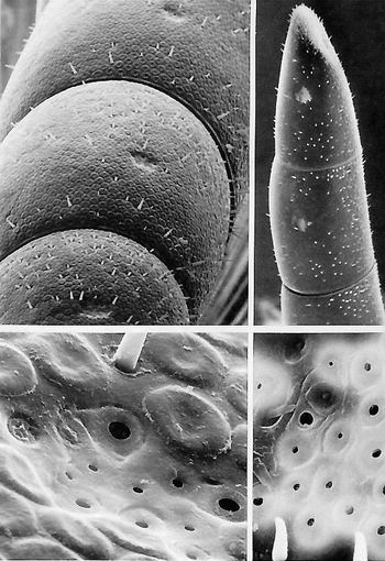
DRONEANTENNATip
TOPRIGHTTerminalthreesegmentsoftheantenna.Atthismagnificationallthatisvisiblearepegorgansandthefaint,pebble-grainedeffectcreatedbytheprofusionofplateorgans.Bothpegandplateorgansareolfactoryorgans.Thepitsonthelasttwosegmentsarereallyaggregationsofahalfdozenorsopitorgans(sensillacoeloconica,sensillaampullacia).(x140)
TOPLEFT.Highermagnificationofthreeantennalsegments.Thethreesenseorgans(peg,pit,andplatesensilla)aremoreevidentatthismagnification.(x420)
BOTTOMLEFT.Tenpitorganssetinacuticulardepressionandsurroundedbyplateorgans.Onesocketedpegisvisibleatthetop.Thepresenceoffreespaceatthepegbasemaypermitthehairshafttomove;ifso,thishairmaybeamechanoreceptor.(x4,200)
BOTTOMRIGHT.Cuticulardepressionontheantenna]segment.Inthisviewpitorganspredominatebuttwo"pits"(at11o'clock)arecapped;theseorgansmaybecampaniformsensilla,andifso,theymustbemechanoreceptorsmonitoringstrainsofthecuticle.Itisnotknownwhythiswholecuticulardepressionappearssobright.Evenifaresidualstaticchargeatthislocusattractedmoregoldintheshadowingprocess,itremainsamysterywhysuchachargewouldbepreferentiallylocatedhereandpersistafterdeath.(x1,705)
DRONEANTENNATIP,VENTRALSURFACE
TOPLowmagnificationoftheantennatip.Atleastfourmorphologicalvariationsofthepegorganscanberesolved,andmostoftheseareclosetotheantennaterminus.Theplateorgansarelocatedmoreposteriorlyandarecircumferentiallyarrangedaroundthissegment(x306)
BOTTOMLEFTHighermagnificationoftheantennashowingthickpegs,plateorgans,andpitorgans.Eachpitorganbearsacuriousbrightmarginarounditsorifice.Odormoleculesenterthepitaseasilyastheypenetratethetinyporesthatradiateoutinspokelikefashionontheplateorgans.(x1,100)
BOTTOMRIGHTThick-setpegorgansandmoreslender,curvedhairs,allsetinsockets.Thecurlingtipsofsomeofthesehairsmaybetheresultofelectronbeamdamage.(x3,300)
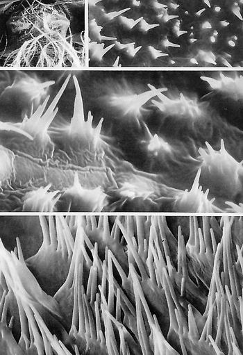
DRONEANTENNASURFACE
BOTTOMRIGHTSurveyoftheterminalsevensegmentsoftheantenna.(x255)
BOTTOMLEFT.Highermagnificationofthetipoftheantennashowingthepacked,ovalplatereceptors(sensillaplacodea)andthewhiskeryappearanceofshortpeg(basiconic)sensillaandslenderhairs(sensillachaetica).Thereareninedistincttypesofsenseorganslocatedontheworkerantenna(onlythreetypesareseeninthisfield).ComparewithPlate2.4,workerantenna.(x60)
TOPHighermagnificationoftheplate,peg,andchaetica-typereceptorsonthepenultimatesegmentoftheantenna.(x1,800)
MIDDLERIGHTPlateandpegorgansonthenexttolastsegmentoftheantenna.Bothtypesareolfactoryorgans.(x3,750)
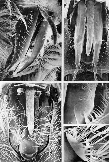
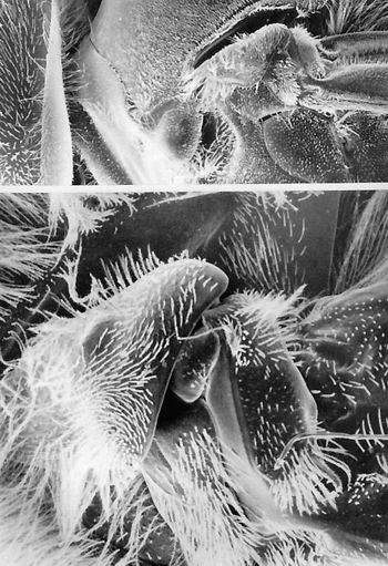
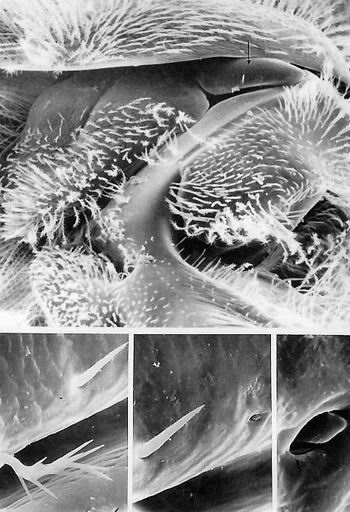
DRONEMOUTHPARTS
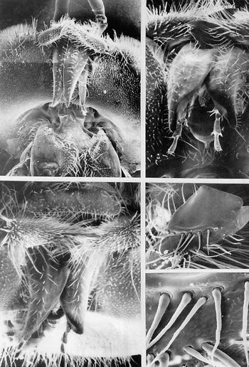
TOPLEFTPhotomontageoftheretractedmouthparts,ventralview.IntheLippercenterofthefieldaretheincisorlike(triangular),pairedgaleae,andtotheimmediaterightisoneofthetwolabialpalps.Crossedmandiblesariseaboveandareatrightanglestothegaleae,withthepairedantennaevisibleabovethemandiblesatthisviewingangle.Theuprighttuftsofsetaeoneithersideofthemandiblesaretheinterfacetalhairsofthecompoundeyes.Thecervixandpairedepisternalscleritesarevisiblebelowthegaleae.ComparewithPlates2.7and2.10,theworkermouthparts.(x31)
TOPRIGHTRetractedmouthparts,frontalview.Atthisviewinganglethecurvedandpointedgaleaepartlyshroudthelabialpaipsandalmostcom-pletelycovertheshortglossa(tongue),whoseterminusisvisibleatthePointwherethegaleaefirstcross.Atthetopofthisfigurearethehirsute,crossedmandibles.(x65)
BOTTOMLEFTHighermagnificationofthefrontalview.Attheverytopofthefieldistheplatelikeclypeus,andsuccessivelybelowthatarethelabrum,mandibles,andgaleae,whichlargelycoverthelabiumwiththeexceptionofthefinalsegmentsoflabialpalps.(x63)
MIDDLERIGHTInteriorsurfaceofthemandibles.Twotiersofstout,socketedhairsareonthismedialsurface.(x135)
BOTTOMRIGHTHighermagnificationofseveralhairsonthemedialsideofthemandibleinthemiddlerightmicrograph.(x450)
DRONEMOUTHPARTS
TOPLEFTDronemouthpartsanteriorview.Thetwodagger-shapedgaleaeoverliethepaired(andsomewhatsplayed-out)labialpalps.Therelativelyshortglossa(tongue)ishiddenfromviewbythesemouthparts.Atthetopofthefieldhirsutemandiblesextendhorizontally,atrightanglestothegaleaeandpalps.(x65)
TOPRIGHT.Close-upofthesurfaceofagaleashowingseveralstout,socketedhairswithflutedsides.Thehairsarisefromthetransverselyfurrowedcuticle.(x1,600)
BOTTOMLEFT.Dronemouthparts,posteriorview.Theveryhirsute,relativelyshort,cylindricalglossahangsdownbetweentheflankinglabialpalps.(x55)
MIDDLERIGHT.Distalfoursegmentsofthelablurn.(x43)
BOTTOMRIGHTLabialpalpwithitsfewpegandtrichoidsensilla.Thetipofthegaleaisimmediatelybelowthelabialpalp.(x170)
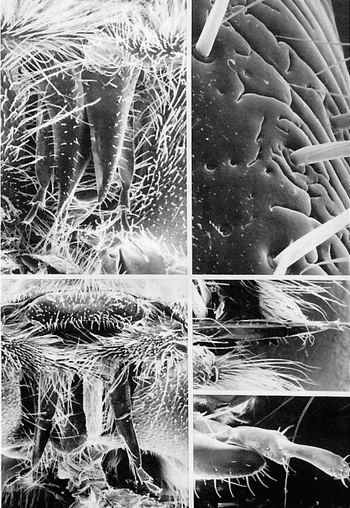
DRONEGLOSSA
TOPLEFT.Glossa(tongue)protrudingoutovertheretractedgaleaeofthemaxillae.Behindthegaleaeandratherobscuredbythemarethepairedlabialpalps.(x63)
TOPRIGHT.Partiallydissectedventralviewoftherelativelyshortglossaflankedbythefour-segmentedlabialpalps.Thelengthofthedroneglossaislessthanhalfthatoftheworker.ComparewithPlate2.7.(x65)
BOTTOMLEFT.Posteriorviewoftheglossa,withfurtherdissectionofthemouthparts(bilateralexcisionofthelabialpalps).Theshortglossahangsdownbetweentherightandleftgaleae,eachwithhairsonthemedialmargin.Betweenthegaleaeandpartlycoveredbytheglossaisthepostmentum.Thearrowpointstoanareafurthermagnifiedinthemiddlerightmicrograph.(x75)
MIDDLERIGHT.Posteriorviewofthebaseoftheglossa.Aglimpseoftheglossaisontheextremeupperleft.Ontherightseveralcuticularhairsofthegalearestonthesideofthepostmentum(arrowinbottomleftmicrograph).(x270)
BOTTOMRIGHT.Distaltipoftheglossa.Theexpanded,spoonliketipiscalledtheflabellum.(x720)
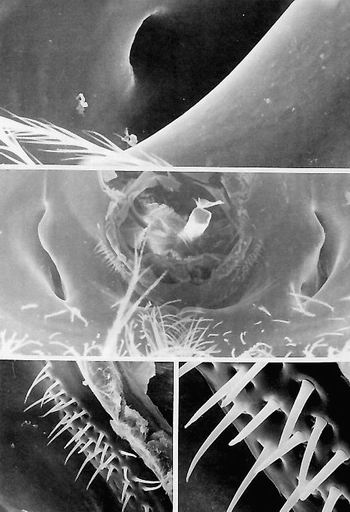
DRONEHEAD,DETACHED,POSTERIORVIEW
MIDDLE.Roundoccipitalforamen,vithsheared-offtissuesprotrudingoutofit,flankedbytwocrescent-shaped,posteriortentorialpits.Twoventrolaterallysituatedhairplatesarelocatedonthecuticularridgeimmediatelyalongsidetheoccipitalforamen.(x144)
BOTTOMLEFT.Entireleft28-setaehairplate.(x550)
BOTTOMRIGHT.Highermagnificationofthelefthairplateshowingthesocketednatureofeachmechanosensoryhair.(x1,760)
TOP.Smallapodemeatthebaseoftherighttentorialpitinthemiddlemicrograph.Therecessleadstotheventralpostoccipitaltentorialarm.(x900)
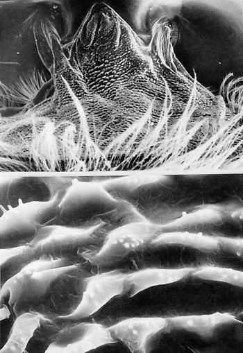
DRONE-CERVIX,DORSALVIEW
TOPDronecervix(neck).Inspecimenpreparation,theheadwasextendedfromthethoraxtostretchoutandshowtheextentofthemembranouscervix.Flexuousbodyhairsfromthepronotumareintheforeground.Thelateralanchoragesforthiscervicalmembranelieabovetheposteriortentorialpits,whiletheinedial(main)insertionisontotherunoftheoccipitalforamen.(x144)
BOTTOMCuticularscalesonthecervixathighermagnification.Eachellipsoidalscaleshowsonetoeightsmalltuberclesprojectingfromitsdorsalsurface.(x2,700)
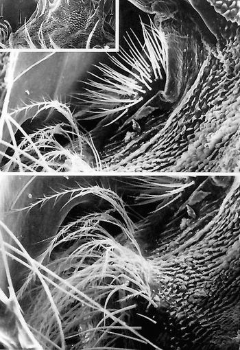
DRONECERVIX
TOPDorsolateralhairplatecontactingthebackofthehead(rightandmiddlearrowsininset).Thesefortytrichoidsensilla,seeminglyinrowsatthisviewingangle,arecervicalinoriginwhilethebranchedhairsarefromthethoracicepisternum.(x288)
TOPINSET.Surveyofthewhole,stretched,dorsalcervix.(x88)
BOTTOM.Dorsolateralcervicalridge(leftarrowininset,topmicrograph)extendingverticallythroughthecenterofthefield.Thelowerextremityofthedorsolateralhairplateisatthetopofthefield(theheadistotheleft).(x360)
DRONECERVIX,VENTRALVIEW
MIDDLEThreetypesof(uninnervated)cuticularornamentation(theposteriorheadistothetop).Thebranchedhairsarefromthethorax;thelow,ellipticalscalesareintheposteriorhalfofthecervix(neck);andthetaller,tufted,multipointedscalesareneartheoccipitalregion.(x144)
TOPOverallcuticularhairdistributionanddensityoftheanteriorcervicaltufts.(x720)
BOTTOMVentrolateralmechanoreceptorhairplateontheoccipitalprocessoftheepisternum(upperleftquadrant).Thecuticular"hole"(farleft)maybepartofanapodeme.(x800)
DRONECERVIX,VENTRALVIEW
TOPLEFTExtendedcervix(neck)(theheadistothetop,thethoraxtothebottom).(X88)
TOPHIGHTCentral"lobe"areaontheventralcervix.CuticularscalesIIIthisareaareforthemostpartsolitaryspinesorblebs.ComparewithPlate2.17,theworkercervix.(x2,200)
MIDDLECervicalmembranewiththecervixextended.Eachcuticularscaleherehasfivetosevenspines,inseverallengths.(x3,600)
BOTTOMCervicalmembranewiththeneckcompressed.Inthisconditionthecuticularspinesfromneighboringscalesmeshandseeminglyoverlap.(x3,600)
DRONETHORAX
Lateralviewofthedronethorax.
DRONEFOREWINGBASE
Top.Photomontageofthedroneforewingbase.Atthefarleftistheheadwiththehairycompoundeye.Atthebottomisthespiracularlobe.Threescleritesareindescendingorderintherighthalfofthefield;mostdorsalisthetriangulartegula,belowwhicharethesupraepimeronandinfraepimeron.Thepleuralsulcusformstheleftsidemarginforthelatterpleurite.Themainveinsoftheleadingedgeofthewingextendtotheright(3o'clock).(x31)
BOTTOM.Close-upofkeywingsclerites.Thetriangulartegulaisthedominantscleriteinthelefthalfofthefield.Totherightofthebasalangleofthetegulaisthebilobedsecondaxillarysclerite.Thisplateisregardedasthepivotalscleriteofthewingbase.Totherightofthesecondaxillaryscleriteisaroughlytrapezoidal(inoutline)scleritecalledthemedianplate.(x100)
DRONEFOREWINGBASE,CLOSE-UP
TOP.Droneforewingbase(theheadistothetop,dorsumtotheright)(seePlate3.18,topmicrograph).Theprobablesensoryreceptorsinthebottomthreemicrographsarelocatedonthepleuralplate(arrow).(x190)
BOTTOMLEFT.Close-upofauniquelybranchedhairandnonsocketedhairaboveonanareacoveredbycuticularscales.(x1,175)
BOTTOMMIDDLE.Hairinthebottomleftmicrograph,withthefieldextendedtoshowthesmallpeglikesensillum.(x1,700)
BOTTOMRIGHT.Highermagnificationofthecuticular"peg"inthebottommiddlemicrograph.Thepegisunusualinthatitisveryflattened,ratherthancolumnar.(x8,500)
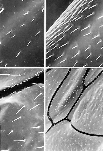
DRONEWING,DORSALSURFACE
BOTTOMRIGHT.Dronewing,dorsalsurface,neartheanteriorwingmarginandwithintheultimatethirdofthewing.Wingveinsshowupasdarkwidelinesmeanderingthroughthewing.(x70)
BOTTOMLEFT.Close-upoftheforewingsurfaceshowingsinglerowsofsocketedsetaeemanatingfromtheveins.Thesemaybemechanoreceptorsinvolvedinmonitoringwingspeed.(x280)湖北天马养蜂场,加我们的微信一起学养蜂。
TOPRIGHT.Leadingedgeoftheforewing.Thesesetae,whichprojectfromtheanteriormostvein,aresocketedandmaypossiblybemechanoreceptors.Thisfieldisanareaabouttwo-thirdsofthedistancetothewingtip.(x650)
TOPLEFT.Veinonthedorsalsurfaceoftheforewingshowingseveralranksofsocketedhairsoneithersideofthewingvein.(x720)PLATE3.20.DRONEWING,DORSALSURFACE